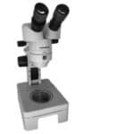
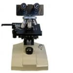
The microscope is an instrument that is used to see the magnified image of objects which are too small for the naked eye. The word microscopic itself denotes a size that can’t be seen without aid. The first account of a microscope can be traced back to thousands of years back in history. From there, it has only been an upward ride full of improvements. In this article, you will get to know the history of the microscope and how it evolved to the way it is today.
Table of Contents
History of the Microscope: The Beginning
The microscope is made from two Greek words- ‘uikpos’ meaning small and ‘okottew’ meaning view.
The first known use of Microscopy is from China. The Chow Foo dynasty invented the Water Microscope. A tube with glasses on both sides had water-filled inside. The level of water varied to achieve different levels of magnification. It is said that the water microscope could achieve 150x magnification. But until now, it is still unproven, Whereas the microscope has become an important part of our life.
Currently, the Microscope has various parts and their functions respectively but its history started with the discovery of glass lenses.
Ancient Romans and Egyptians have claimed to have certain glass pieces that made objects look larger. The Greeks were also running parallel in the race. Someone then discovered a crystal that could set fire to cloth or paper using Sun rays. This led to the crystal being called Burning glasses. This term is mentioned by Roman philosophers in their writings in the first century AD.
The use of these crystals remained uncommon until the 13th century. These crystals came to be known as lenses as their shape resembled the seed of lentils. The earliest known microscope in the Roman empire was a tube with a plate and lens fitted on the other side. People would keep the object on the plate and observe from the other side. The magnification was lesser than 10x. The locals started calling this as Flea Glasses.
The invention of spectacles is credited to two people in Tuscany, Italy in the 14th century. Salvano d’Aramento degli Amati and Allessandro della Spina both independently developed spectacles. But both of them kept the process a secret. With many people working on glasses, the use of spectacles was common in the 15-16th century.
Compound Microscopes
Around 1590, a Dutch pair of father and son, Zaccharias Janssen and Hans Jenssen along with their neighbor Hans Lippershey worked on glasses. The special maker pair experimented with various lenses and their combinations. They discovered that they could produce enlarged images of small. This is said to be the invention of the compound microscope. Another important point they learned was that with multiple lenses, the image could be magnified further.
The microscope had an eyepiece and an objective lens. The instrument was made of cardboard and wood. In the 17th century, English philosopher Robert Hooke used a microscope for giving his study. He was also used for his demonstrations to the Royal Society. In 1665 he published Micrographia where he coined the word cell. In the book, he also mentioned how the lens could prepare a serviceable microscope.
During this time Galileo also worked on microscopes.
He experimented, pointed out the principles, and made lots of improvements in the existing design.
The biggest break-through was by Anton van Leeuwenhoek. He worked at the same time as Robert Hooke, 17th century. He is known as the Father of Microscopy. He worked in a dry goods store where he used glasses to count threads. He was his own teacher. He learned new methods of polishing and grinding lenses of various curvatures.
His work led to a magnification of 270x. It was the highest at that time. All this led to the discovery of microscopic creatures. He became the first person to observe bacteria, drops of water, yeast etcetera. He wrote a lot of letters to societies of England and France about his discoveries. His microscope could give details of around 0.7 micrometers.
Robert Hooke later confirmed his theories and made his own copy of the microscope with some changes. Hooke’s version had a diaphragm, balance springs, and other improvements. Some of these improvements are still present in the modern-day microscope. Robert Brown used this microscope and discovered the Nucleus in 1831.
The 18th And 19th Century
The next major step took place around 100 years later. This milestone was Charles Hall inventing the achromatic lens in the 1730s. This solved the problems by Chromatic aberration. Due to single lenses, the microscope would produce spurious colors.
These aberrations were even worse in compound microscopes where more than one lens was used. The microscopes would produce inferior images. Being an optician, Hall, tried many combinations of lenses to prevent the aberrations. He finally succeeded with a combination of concave flint and a convex crown glass lens. It proved that the use of a second lens with different shape and refracting properties could prevent color distortion.
Benjamin Martin in 1774 produced a color-corrected lens for a microscope.
Joseph Lister in 1830 found a cure to the problem of Spherical aberration. He placed the lens at a certain distance from each other. Both Charles Hall and Joseph Lester improved the picture quality of the microscope. Previously, the quality was inferior to distorted images.
Up to Joseph Lister, all the improvements were for the betterment of the image quality.
In 1863, the Ernst Leitz company found a cure for a mechanical problem. They introduced a revolving turret that had 5 objectives.
The next major improvement was Abbe Condenser. It was made by Carl Zeiss and Ernst Abbe at Zeiss Optical works. Abbe laid out the approach of computational optics development. He differentiated between the resolution and magnification of the image. His work resulted in the Abbe condenser. Abbe’s work led to the development of 17 microscope objectives. All the objectives were based on mathematical modeling. He carefully studied the materials, designs, curvature, aperture etcetera.
From this point onwards, the design started to be on the laws of physics. The trial and error approach was excluded and criticized. Many companies started manufacturing plants for more precise microscopes. Lots of investment in Research and development resulted in many improvements. One such was by another Zeiss employee, August Kohler. In 1893, Kohler used double diaphragms and created an illumination system that was uniform. He succeeded in getting a bright image with minimum glare. This is known as Kolher Illumination.
At this time the market for microscopes grew very large.
In 1904, Zeiss introduced the first UV microscope. It had double the resolution of a visible light microscope. Further developments came as the discovery of phase contrasts and differential interference contrast. The discoveries were made by Frits Zernike in 1953 and Georges Nomarski in 1955 respectively. Both these developments lead to unstained and transparent samples.
Electron Microscope
German scientists Max Knoll and Ernst Ruska in 1931 invented the electron microscope. This microscope exceeded the limits of light. Light microscopes could produce magnification up to 500x or 1000x. The electron microscope achieved a magnification of 2million times. Knoll and Ruska worked on a transmission electron microscope (TEM). Later in 1942 Ruska built the scanning electron microscope (SEM).
Fluorescent Microscopes
They are the latest inventions in the light microscope industry. The main techniques are chemical staining, use of antibodies etcetera. The development of fluorescent microscopes led to a major design- confocal microscopes.
Advanced light sources like Halogen, LED have helped improve the light microscope.
Super-Resolution Microscopes
There are current projects of the optical microscope industry. It focuses on the super resolution. They use Stimulated emission depletion (STED) microscopy. The STED was developed by Stefan Hell for which he got a Nobel prize.
Digital Microscope
Digital microscopes have revolutionized the microphotography. They integrate a microscope camera on the port of a microscope. This allows the live image transmission into a screen.
Dino-Lito Microscope
A 21st-century invention, these are hand-held microscopes. They are very compact with a size comparable to a pen. They offer magnification up to 500x but with slightly low zoom quality.
X-Ray Microscope
These microscopes use electromagnetic radiation to image objects. They can produce 3-D images. They can also produce images of chemically unfixed biological materials. X-ray microscopes are currently research.
Best Microscopes In The Market
1. Swift Compound Monocular Microscope SW200DL
- 【MAGNIFICATION SETTING】Available magnification settings of 40X, 100X, 250X, 400X, and 1000X through aberration-correcting 4X, 10X, and 40X glass objectives with wide-field 10X and 25X eyepieces
- 【Dual lllumination】Dual illumination system allows you to examine both transparent and solid specimens; cool LED lights protect eyesight and live specimens
- 【Excellent material】Rugged design with metal arm and base, carrying handle, and cordless capability make this compound microscope a practical pick for field experiments
This Swift SW200DL microscope is portable and suitable for both students and hobbyists. It has a solid frame and a sturdy metal body. The 2 in 1 illumination system provides strong and powerful microscopy. The magnification range is 40x to 1000x. It has three aberration correcting glasses. This gives a blur-free image. The head is fixed at a 45-degree angle for preventing the neck strain. There are three objective lenses- 4x,10x, and 40x. The cool LED light protects your eyesight.
2. ESAW MM02 Medical Microscope
- High quality 10xWF and 15xWF EYEPIECE
- Magnification 100x TO 1500x
- 1 year Manuafcturer's warranty but no breakage included.
ESAW is a common name for microscopes in school and scientific labs. It is suitable for academics in school and at home. The microscope has an LED substage. It comes with 50 already prepared slides that cover many topics. It can magnify up to 1500x. There are 5 magnification settings- 100x, 150x. 450x, 675x, 1000x, and 1500x. You also get a cleaning cloth and dust cover. The product is easy to use with coarse and fine motion knobs.
3. Gemkolabwell GS701-00 Microscope
This Gemkolabwell microscope is suitable for students and academicians. It has a 10x objective, 1000x magnification, and 10x EP. It has a light-reflecting mirror. This mirror helps in working with an external light source. You also get some prepared slides with the microscope. Anti-glare, achromatic anti-fungus, and hard coating are extra advantages of this product.
Final Thought
We have tried to give you an idea about the history of the microscope in brief. All the major events related to microscope is covered. If the microscope is a part of your daily life, you should definitely check out how you can maintain one. We think it is more than enough if you want to learn about it.


Leave a Reply